| Reference: | P-24-U:2002-208, LA-UR-04-1474 |
|---|---|
| From: | Damian Swift, P-24 |
| To: | Distribution |
| Date: | 9 Aug 02; revised 27 Aug and 12 Sep 02 |

Opportunistic:
| TRIDENT schedule | |||
|---|---|---|---|
| Plan | Actual | Comments | |
| Start of set-up | 26 Jul | 28 Jul | Conflict with LLNL TXD meeting |
| Start of facility time | 29 Jul | 29 Jul | |
| First shot | 30 Jul | 30 Jul | Actual: later in day than planned. |
| End of facility time | 8 Aug | 8 Aug | S target area left set-up |
| Shot allocations | |||
|---|---|---|---|
| Plan | Actual | Comments | |
| Timing etc | ~5 | 2 | Actual excludes some Si recovery |
| NiTi LDRD | 20-30 | 29 | Used all but 2 available. |
| NiAl LDRD | 6-10 | 7 | Used all provided. |
| Ellipsometry | 10-20 | 17 | Includes Sn and Si calibration shots. |
| Si recovery | 0 | 6 | Excludes calibration shots for ellipsometry. |
| Shear-banding | 0 | 2 | |
| Total | 40-50 | 63 | |
| Firing duration and rate | |||
|---|---|---|---|
| Plan | Actual | Comments | |
| Experiment time | 6.5 | 5.5 | Unanticipated delays and down-time. |
| Shots/day | 7.0 | 11.5 | Higher shot rate at low energy. |
| NiTi 3D | nominal 50.0% Ni, from Bob Hackenberg (MST-6) |
| NiTi 5D | nominal 50.4% Ni, from Bob Hackenberg (MST-6) |
| NiAl | from Ken McClellan and John Brooks, MST-8 |
| Si (100) | phosphorus-doped Goodfellow |

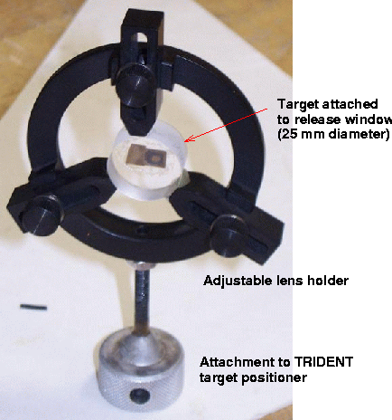
The NiAl samples were prepared by polishing after gluing to a window. The only windows available in the necessary time scale were too large to be held in the clamp-type holder. A holder was constructed based on a lens holder, with three radially-sliding bars to grip the sample. It was found necessary to use a little "tacky wax" to secure the sample to the holder and eliminate any propensity for the surfaces to slip.

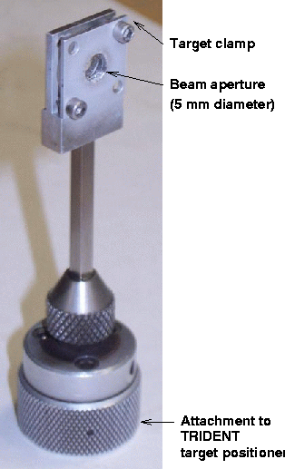
The Fresnel zone plate was used to smooth the beam spatially. With the unsmoothed beam at best focus, inserting the phase plate gave a focal spot nominally 4 mm in diameter. However, the undiffracted light produced a hotspot in the centre of the disc, so the usual procedure was followed of defocusing the drive beam by 3/4". The resulting spot was ~5 mm in diameter, and was estimated to contain 85% of the nominal drive energy reported for each shot.
In the VISAR `shots the target was close to normal to the drive beam. In the ellipsometry shots, the target was rotated significantly, so the intensity of the drive beam was lower.

timingXXus.ipl.
| sweep speed | start | from 14910 | from 14922 | from 14948 |
|---|---|---|---|---|
| (ns) | ||||
| 50 | 2962 | 2340 | 2190 | 2390 |
| 20 | 3002 | 2230 | 2430 | |
| 10 | 3013 | 2241 | 2441 | |
| 5 | 3023 | 2251 | 2451 | |
| camera from laser | - | 499 | 649 | 449 |
Using the fiducial markers, the camera scale and nominal offset were as follows:
| sweep speed | temporal scale | location of drive pulse | first fiducial pulse | ||
|---|---|---|---|---|---|
| (ns) | (ps/pixel) | (pixels from start of record) | (ns from start of record) | (pixels from start of record) | (ns from start of record) |
| 50 | 41.4±0.5 | 502 | 20.76±0.25 | 400 | 16.54±0.20 |
| 20 | 18.1±0.1 | 496 | 8.96±0.10 | 266 | 4.81±0.05 |
(The location of the drive pulse is with the delay set to the value for the drive to occur nominally at the centre of the record.)
For a given value of the delay d, reference delay dr and drive pulse time p on the reference record, times in the record are expressed with respect to the drive pulse by setting the the record to start at d - dr - p. Alternatively, if the Nth fiducial pulse occurs at a time f in the record compared with a time fr in the reference record, times in the record are expressed with respect to the drive pulse by setting the the record to start at fr - (p + f) + (N - 1) tp, where tp is the interval between fiducial pulses.
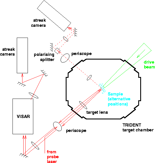
The VISAR probe laser was used as the source, with the omission of the cylindrical lens used to illuminate a line on the sample and the addition of a depolariser at the same point in the system. The angles of incidence and reflection were 32±0.5o to the sample normal. The target was rotated to send specular reflections from the probe laser towards the detecting optics; thus as designed it was not possible to use VISAR and ellipsometry with specular reflection on the same experiment in this initial scoping study. Light reflected from the sample was collected with a 200 mm f/5 achromat positioned with the sample at its focal length. A polarising cube was used to split the two components, and these were recorded using a second Hamamatsu streak camera, with an S-1 photocathode.
The two channels were focused onto the camera slit through the same lens (200 mm f/5 achromat); the resulting beams diverged too far to be captured by the camera so a piece of tape was stuck over the slit to act as a diffuser.
| Shot | Target | Line VISAR | Ellipsometer | Pulse | Energy | Comments | |||
|---|---|---|---|---|---|---|---|---|---|
| sweep | delay | sweep | delay | (ns) | standard | LP: raw | |||
| (ns) | (ns) | (ns) | (ns) | (J) | (mJ) | ||||
| 30 Jul | |||||||||
| 14906 | Al/Si (100), 30 µm | 50 | 2982 | - | - | 2.5? | 20±5 | - | omitted camera filter: swamped by stray drive light |
| 14907 | Al/Si (100), 30 µm | 50 | 2982 | - | - | 2.5? | 20±5 | - | very fast jumpoff |
| 31 Jul | |||||||||
| 14909 | NiTi 3D, 90 µm (#1) | 50 | 2982 | - | - | 2.5 | 20±1 | - | |
| 14910 | NiTi 3D, 198 µm (#10) | 50 | 2380 | - | - | 2.5 | 18±1 | - | timing offset changes |
| 14911 | NiTi 3D, 400 µm (#13) | 50 | 2430 | - | - | 2.5 | 21±1 | - | |
| 14912 | NiTi 3D, 99 µm (#2) | 50 | 2355 | - | - | 1.0 | 21±1 | - | no VISAR |
| 14913 | NiTi 3D, 101 µm (#4) | 50 | 2355 | - | - | 1.0 | 23±1 | - | |
| 14914 | NiTi 3D, 210 µm (#12) | 50 | 2377 | - | - | 1.0 | 27±1 | - | |
| 14915 | NiTi 3D, 408 µm (#14) | 50 | 2420 | - | - | 1.0 | 33±1 | - | |
| 14916 | NiTi 3D, 95 µm (#3) | 50 | 2360 | - | - | 1.0 | 6±1 | - | |
| 14917 | NiTi 3D, 182 µm (#11) | 50 | 2380 | - | - | 1.0 | 7±3 | - | |
| 14918 | NiTi 3D, 397 µm (#15) | 50 | 2420 | - | - | 1.0 | 8±3 | - | |
| 14919 | Si (100), 380 µm | 50 | 2400 | - | - | 1.0 | 28±1 | - | sample recovered |
| 1 Aug | |||||||||
| 14921 | Si (100), 380 µm | 50 | 2400 | - | - | 1.0 | 28±1 | - | poor probe level; sample recovered |
| 14922 | Si (100), 380 µm | 50 | 2250 | - | - | 1.0 | 27? | - | new timing offset; poor probe level; sample recovered |
| 14923 | Si (100), 380 µm | 50 | 2250 | - | - | 1.0 | 15±1 | 0.820 | sample not recovered |
| 14924 | NiTi 3D, 91 µm (#5) | 50 | 2210 | - | - | 1.0 | 15±1 | 0.761 | |
| 14925 | NiTi 3D, 103 µm (#9) | 50 | 2200 | - | - | 1.0 | ~4 | 0.0512 | |
| 14926 | NiTi 3D, 89 µm (#7) | 50 | 2200 | - | - | 0.4 | ~3 | 0.1308 | |
| 14927 | NiTi 3D, 87 µm (#8) | 50 | 2200 | - | - | 0.4 | - | 0.0418 | |
| 14928 | NiTi 3D, 105 µm (#6) | 50 | 2200 | - | - | 2.5 | - | - | energy to be deduced from photodiode record, cf previous shot |
| 14929 | Si (100), 380 µm | 50 | 2260 | - | - | 2.5 | 13 | 0.651 | missed shock; recovered sample |
| 14930 | Si (100), 380 µm | 50 | 2240 | - | - | 2.5 | 17 | 0.825 | sample in small pieces |
| 14931 | Al/Si (100), 30 µm/LiF, 2 mm | 20 | 2230 | - | - | 2.5? | - | 0.621 | recovered sample |
| 5 Aug | |||||||||
| 14934 | Al/Si (100), 30 µm/LiF, 2 mm | 20 | 2230 | - | - | 1.0? | 27 | 1.360 | recovered sample |
| 14935 | NiTi 5D, 105 µm (#1) | 50 | 2200 | - | - | 1.0 | 28 | 1.428 | probe intensity low but usable |
| 14936 | NiTi 5D, 194 µm (#6) | 50 | 2240 | - | - | 1.0 | 29 | 1.354 | |
| 14937 | NiTi 5D, 408 µm (#11) | 50 | 2280 | - | - | 1.0 | 24 | 1.298 | |
| 14938 | NiTi 5D, 101 µm (#3) | 50 | 2210 | - | - | 1.0 | ~17 | 0.565 | |
| 14939 | NiTi 5D, 200 µm (#7) | 50 | 2240 | - | - | 1.0 | 13 | 0.614 | |
| 14940 | NiTi 5D, 409 µm (#12) | 50 | 2280 | - | - | 1.0 | 8 | 0.549 | |
| 14941 | NiTi 5D, 87 µm (#2) | 50 | 2210 | - | - | 1.0 | 8 | 0.437 | |
| 14942 | NiTi 5D, 196 µm (#8) | 50 | 2240 | - | - | 1.0 | 6 | 0.393 | |
| 6 Aug | |||||||||
| 14944 | NiTi 5D, 399 µm (#13) | 50 | 2270 | - | - | 1.0 | 10 | 0.449 | no probe signal |
| 14945 | NiTi 5D, 99 µm (#4) | 50 | 2210 | - | - | 1.0 | - | 0.0557 | |
| 14946 | NiTi 5D, 185 µm (#9) | 50 | 2230 | - | - | 1.0 | - | 0.0537 | probe signal weak but usable |
| 14947 | NiTi 5D, 402 µm (#14) | 50 | 2270 | - | - | 1.0 | - | 0.0427 | probe multi-moded; signal weak but may be usable |
| 14948 | NiTi 5D, 394 µm (#15) | 50 | 2470 | - | - | 2.5? | -? | 0.566? | new VISAR timings |
| 14949 | NiTi 5D, 200 µm (#10) | 50 | 2430 | - | - | 0.4 | 5 | 0.216 | |
| 14950 | NiAl (110)+(111), ~120 µm / BK7 (P2) | 50 | 2410 | - | - | 1.0 | 33 | 1.509 | |
| 14951 | NiAl (110)+(111), 149 µm / BK7 (P3) | 50 | 2420 | - | - | 1.0 | 25 | 1.198 | probe laser multi-moded but some fringe motion visible |
| 14952 | NiAl (110)+(111), 122 µm / BK7 (P1) | 50 | 2415 | - | - | 1.0 | 32 | 1.293 | little fringe motion (glue?) but evidence of side-to-side variation |
| 14953 | NiAl (110)+(111), 84 µm / BK7 (P4) | 50 | 2415 | - | - | 1.0 | 29 | 1.365 | forgot filter: fringes obscured by scattered green |
| 14954 | NiAl (100)+(111), 185 µm / BK7 (P9) | 50 | 2410 | - | - | 1.0 | 27 | 1.308 | fringes moved one side only |
| 7 Aug | |||||||||
| 14956 | LLNL Al/groove/wax (B) | - | - | - | - | 0.8 | ~180 | - | no calorimeter reading |
| 14958 | LLNL Al/groove/wax (A) | - | - | - | - | 2.5 | 158 | - | photodiode scale same as 14958 except extra OD 0.3: use to deduce 14956's energy |
| 14959 | Sn, 25 µm | 20 | 2435 | - | - | 1.0 | 27 | 1.376 | Focus preshot for scale: ~5 mm dia holder. Probe multimoded; sample destroyed. |
| 14960 | Sn, 25 µm | 20 | 2435 | - | - | 2.5 | 25 | 1.205 | |
| 14961 | Sn, 25 µm | 20 | 2435 | - | - | 2.5 | ~2? | - | |
| 14962 | Sn, 25 µm | 20 | 2438 | - | - | 2.5 | 13 | 0.497 | |
| 14963 | Sn, 25 µm | 20 | 2438 | - | - | 2.5 | - | 0.112 | |
| 14964 | Sn, 25 µm | 20 | 2438 | - | - | 2.5 | 19 | 0.848 | |
| 14965 | Sn, 25 µm/LiF | 20 | 2438 | - | - | 2.5 | 13 | 0.425 | |
| 8 Aug | |||||||||
| 14967 | Al/Si (100), 30 µm/LiF | - | - | 50 | 2972 | 2.5 | 33 | 1.577 | omitted camera filter: swamped by stray drive light |
| 14968 | Al/Si (100), 30 µm/LiF | - | - | 50 | 2972 | 2.5 | 54 | 2.55 | saw effect |
| 14969 | Al/Si (100), 30 µm/LiF | - | - | 50 | 2972 | 2.5 | 88 | 4.27 | saw effect |
| 14970 | Sn, 25 µm/LiF | 20 | 2438 | 50 | 2972 | 2.5 | 9 | 0.428 | no effect (?); diffuse VISAR signal |
| 14971 | Sn, 25 µm/LiF | 20 | 2438 | 50 | 2972 | 2.5 | 40 | 2.279 | no effect (?) |
| 14972 | Sn, 25 µm/LiF | 20 | 2438 | 50 | 2972 | 1.0 | 54 | 2.77 | saw effect |
| 14973 | Sn, 25 µm | 20 | 2438 | 50 | 2972 | 1.0 | 55 | 2.90 | VISAR washed out; ellipsometer not useful. |
| 14974 | NiAl (100)+(111), 203 µm / BK7 (P10) | 50 | 2424 | - | - | 2.5 | 8 | 0.625 | spatial variation in fringe motion |
| 14975 | NiAl (110)+(111), 145 µm / BK7 (P7) | 50 | 2414 | - | - | 2.5 | 18 | 0.767 | spatial variation in fringe motion |
| 14976 | Sn, 25 µm/LiF | 20 | 2438 | - | - | 2.5 | 10? | 0.829 | Ramp wave. Forgot to replace filter: fringes washed out by stray drive light. |
15 samples of alloy 3D and 14 samples of alloy 5D were shocked and recovered; intensity not exactly as specified for each but within accuracy found possible in practice. One VISAR record lost; pressure pulse captured on all other shots (despite long transit time for the thick samples). One extra sample of each composition was not fired, as the shots already obtained were deemed to cover the pressure range planned, and time was needed to compensate for laser outage.
Problems were experienced with glue joints between the samples and the BK7 release window, and the alignment of the interface may have been poor on early shots. Spatial variations were seen clearly in later shots, with some evidence in early shots.
Time-varying differences in the absolute and relative intensity from each channel was observed clearly in the experiments on Si and in the higher-pressure experiments on Sn. Pressure-dependent birefringence of LiF may contribute to the observed effects. Complementary experiments to calibrate the response of Sn to laser-induced loading produced VISAR records of reasonable quality, though they did not cover the higher intensity range where strong ellipsometry effects were observed.
Secondary objectives:
| Bob Hackenberg | NiTi samples and planning |
| John Brooks and Ken McClellan | NiAl samples and planning |
| Jose Velarde | manufacture of clamp-type target holders |
| Robi Mulford | elephant design and manufacture |
| Johnathan Niemczura | target assembly and positioning, optics work |
| Randy Johnson | Laser/optics consulting, VISAR alignment, TLC for probe laser |
| Tom Hurry, Nathan Okamoto, Fred Archuleta, Ray Gonzales | target area and laser work |
| Dennis Paisley | discussions of laser flyers with visitors |
| Dan Thoma | PI for NiTi LDRD |
| Aaron Koskelo | PI for NiAl LDRD |
| Allan Hauer | Project support (use of TRIDENT) - C10: HEDP |
| Cris Barnes | cbarnes@lanl.gov
|
| Steve Batha | sbatha@lanl.gov
|
| John Brooks | jdbrooks@lanl.gov
|
| Juan Fernández | juanc@lanl.gov
|
| Robert Gibson | rbg@lanl.gov
|
| Robert Hackenberg | roberth@lanl.gov
|
| Allan Hauer | hauer@lanl.gov
|
| Randy Johnson | rpjohnson@lanl.gov
|
| Aaron Koskelo | koskelo@lanl.gov
|
| George Kyrala | kyrala@lanl.gov
|
| Ken McClellan | kmcclellan@lanl.gov
|
| Carter Munson | cmunson@lanl.gov
|
| Johnathan Niemczura | doc@lanl.gov
|
| Dennis Paisley | paisley@lanl.gov
|
| Damian Swift | dswift@lanl.gov
|
| Dan Thoma | thoma@lanl.gov
|
| External: | |
|---|---|
| UCSD (Bimal Kad) | |
| ASU (Pedro Peralta) | |
| LLNL (Dan Kalantar, Jim McNaney) | |
| Oxford U (Justin Wark, Jim Hawreliak) | |